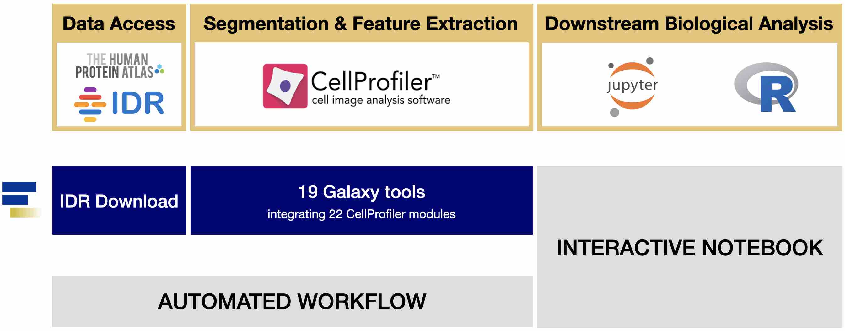

Data acquisition protocol and quality control Integration of this dataset with datasets resulting from other types of perturbations (e.g., patient cell samples or genetically perturbed samples) enables identification of small-molecule mimetics of particular disease- or gene-related phenotypes that could be useful as probes or potential starting points for development of future potential therapeutics. A dataset comprising small-molecule perturbations, as presented here, can be used for small-molecule library enrichment (to create smaller libraries while retaining high diversity of phenotypic impact) and small-molecule mechanism-of-action studies, including target identification.

The applications of image-based profiling are many and diverse.

Further analysis then aggregates the data into multivariate profiles of these features to compare signatures among sample treatments.
#Cellprofiler 2.2.0 software#
The “Cell Painting” assay used for the dataset presented here uses fluorescent markers to broadly stain a number of cellular structures in high-throughput format, while automated software extracts the single-cell image-based morphological features. In contrast to a screening strategy, where a usually limited number of features are quantified to select for a known cellular phenotype, profiling relies on collecting a large suite of per-cell morphological features and then using statistical analysis to uncover subtle morphological patterns (“signatures”) by which the perturbations can be characterized. Phenotypic profiling has emerged as a powerful tool to discern subtle differences among treated samples in a relatively unbiased manner. In this way, the morphological characteristics (or “phenotype”) of cells, tissues, or even whole organisms can be examined, along with the concomitant changes induced by the perturbants of choice. Ī typical imaging assay uses several fluorescent probes (or fluorescently tagged proteins) simultaneously with stain cells, each labeling distinct cellular components in each sample. Concurrently, the advent of high-throughput imaging has also become an engine for pharmacological screening and basic research by allowing multiparametric image-based interrogation of physiological processes at a large scale. While microscopy has enriched our understanding of biology for centuries, only recently has robotic sample preparation and microscopy equipment become widely available, together with large libraries of chemical and genetic perturbations. High-throughput quantitative analysis of cellular image data has led to critical insights across many fields in biology.


 0 kommentar(er)
